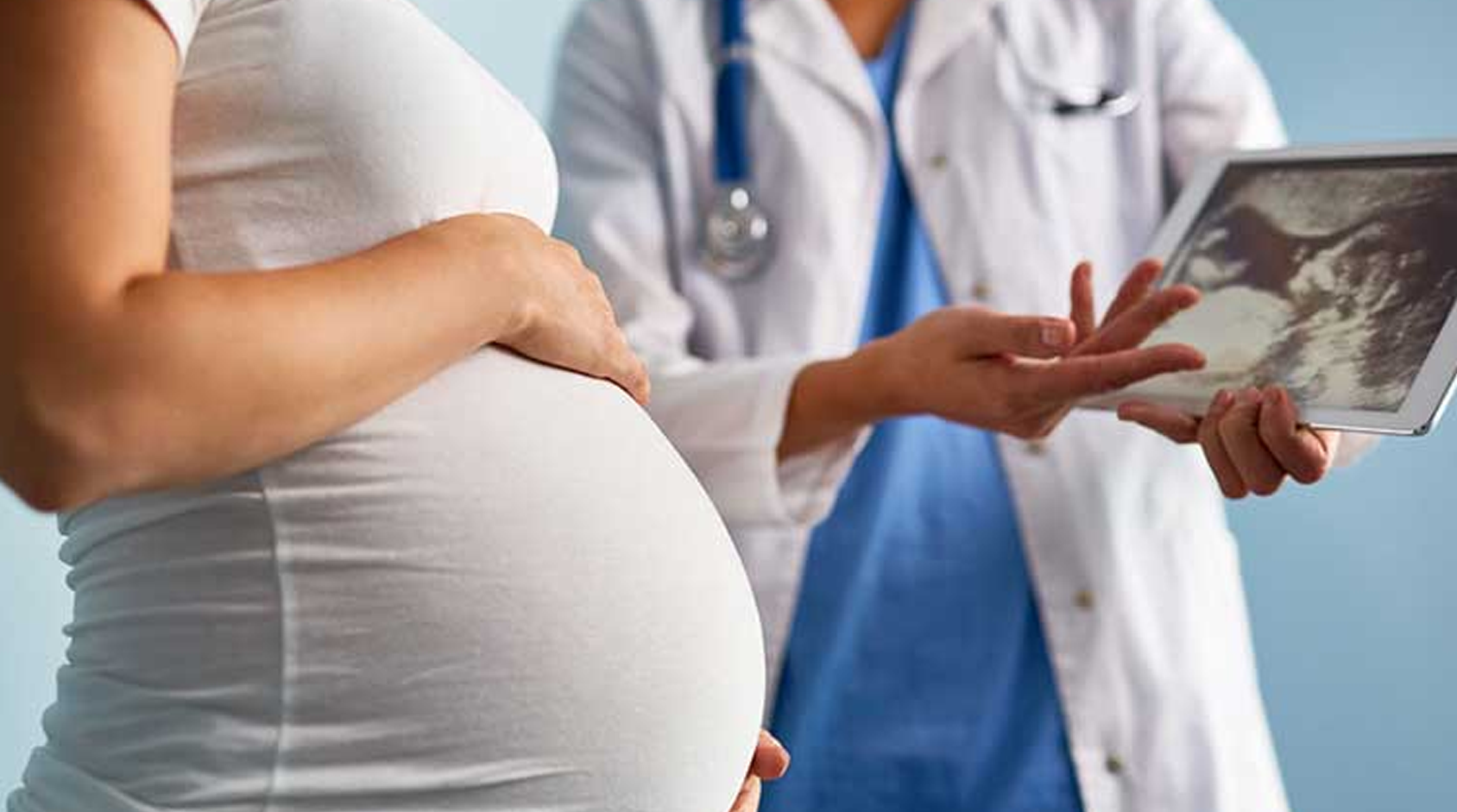
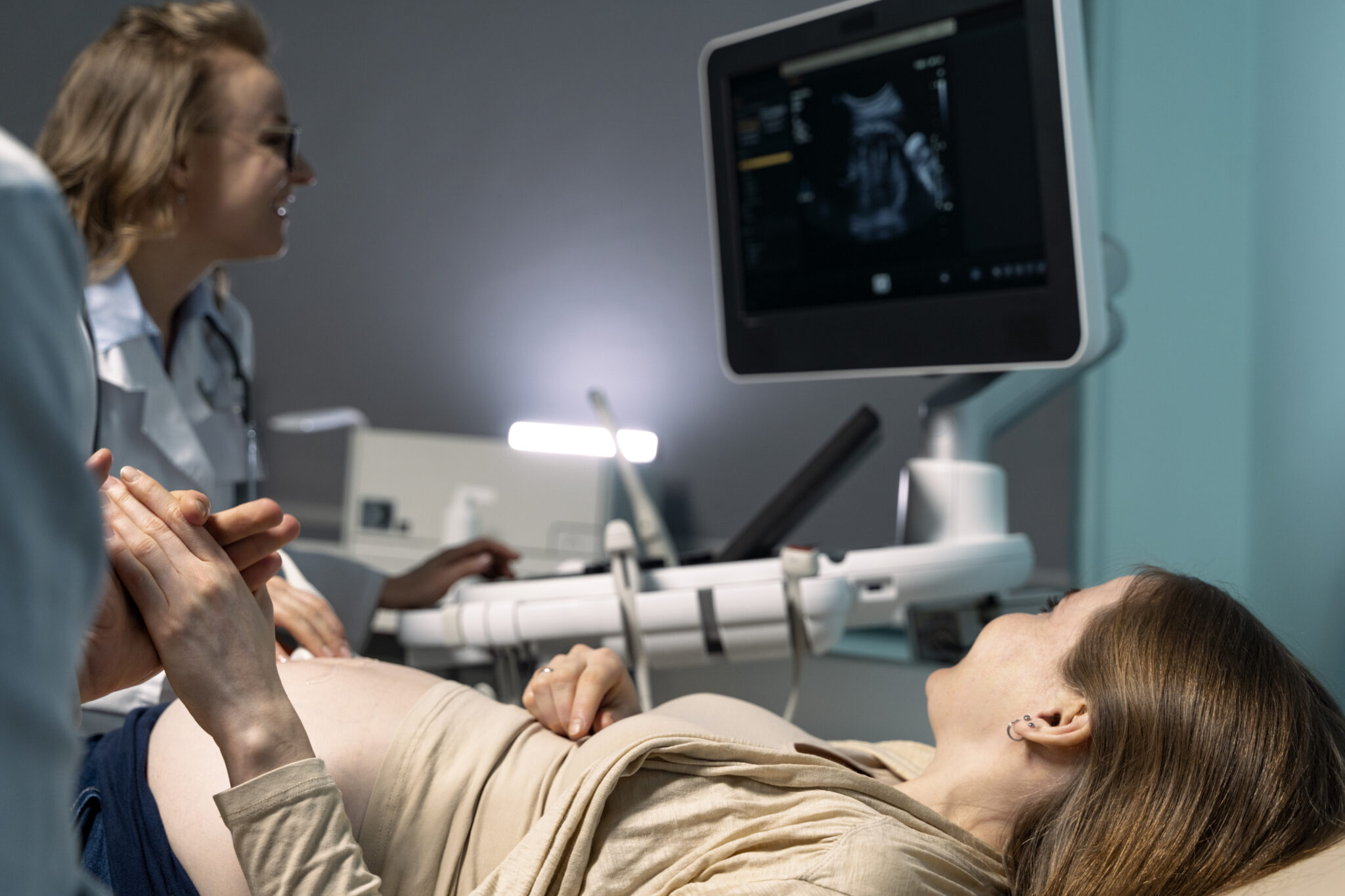
Ultrasound screening in pregnancy involves using high-frequency sound waves to create images of the fetus, uterus, placenta, and surrounding structures in the mother’s abdomen and pelvis. It’s a non-invasive and safe procedure that provides valuable information about the developing fetus and the progress of the pregnancy.
An ultrasound, also called a sonogram, can help monitor normal fetal development and screen for any potential problems. Along with a standard ultrasound, there are a few more advanced ultrasounds — including a 3-D ultrasound, a 4-D ultrasound, and a fetal echocardiography, which is an ultrasound that looks in detail at the fetus’ heart.
First trimester NT scan
The first-trimester nuchal translucency (NT) scan is a specialized ultrasound screening performed between 11 and 14 weeks of pregnancy. It aims to assess the risk of chromosomal abnormalities, particularly Down syndrome (trisomy 21), as well as other conditions such as Edwards syndrome (trisomy 18) and Patau syndrome (trisomy 13).
During the NT scan:
- Measurement of Nuchal Translucency: The thickness of the fluid-filled space at the back of the fetus’s neck is measured using ultrasound. An abnormally thickened nuchal translucency may indicate an increased risk of chromosomal abnormalities or certain structural defects.
- Crown-Rump Length (CRL): The length of the fetus from crown (top of the head) to rump (bottom) is measured. This measurement helps determine the gestational age and ensures accurate dating of the pregnancy.
- Assessment of Nasal Bone: The presence or absence of the nasal bone is evaluated during the scan. The absence of a nasal bone can be associated with an increased risk of chromosomal abnormalities, particularly Down syndrome.
- Blood Test: In conjunction with the NT scan, a blood test known as the first-trimester combined screening test may be performed. This blood test measures the levels of certain proteins and hormones in the mother’s blood, including pregnancy-associated plasma protein-A (PAPP-A) and human chorionic gonadotropin (hCG). Abnormal levels of these substances may further indicate an increased risk of chromosomal abnormalities.
- Risk Assessment: The measurements from the NT scan, along with maternal age and the results of the blood test, are used to calculate the risk of chromosomal abnormalities. This risk assessment helps guide further testing or counseling for the expectant parents.
It’s important to note that the NT scan is a screening test, not a diagnostic test. A positive result does not confirm the presence of a chromosomal abnormality but rather indicates an increased risk, warranting further testing such as chorionic villus sampling (CVS) or amniocentesis for definitive diagnosis.
The first-trimester NT scan provides valuable information for expectant parents, allowing them to make informed decisions about additional testing and to prepare for any potential outcomes. It also offers reassurance to many families when results indicate a low risk of chromosomal abnormalities.
2nd trimester anatomy scan
The second-trimester anatomy scan, also known as a fetal anomaly scan or mid-pregnancy ultrasound, is typically performed between 18 and 22 weeks of gestation. This comprehensive ultrasound examination provides a detailed assessment of the fetus’s anatomy and development, offering valuable information to both healthcare providers and expectant parents.
During the second-trimester anatomy scan:
- Fetal Anatomy Evaluation: The ultrasound technician or obstetrician carefully examines various structures and organs of the fetus, including the brain, spine, heart, lungs, stomach, kidneys, bladder, limbs, and umbilical cord. This thorough assessment helps identify any structural abnormalities or congenital anomalies.
- Measurement of Fetal Growth: The technician measures the fetus’s head circumference, abdominal circumference, and femur length to assess growth and ensure that the fetus is developing appropriately for its gestational age.
- Placental Location and Health: The ultrasound evaluates the position, size, and structure of the placenta, which plays a crucial role in supplying nutrients and oxygen to the fetus. Abnormalities such as placenta previa or placental insufficiency may be detected during the scan.
- Amniotic Fluid Assessment: The technician examines the volume and distribution of amniotic fluid surrounding the fetus. Too much or too little amniotic fluid can indicate potential issues such as fetal distress or certain birth defects.
- Fetal Position: The technician notes the fetal position within the uterus, which can provide information about the likelihood of a breech presentation or other positioning issues that may impact delivery.
- Gender Determination: In many cases, the second-trimester anatomy scan offers the opportunity for parents to find out the baby’s gender if they choose to know.
The second-trimester anatomy scan is a crucial component of prenatal care, as it helps identify any potential fetal abnormalities or complications early in pregnancy. Early detection allows for appropriate medical management, counseling, and planning for the birth and care of the baby. Additionally, the ultrasound provides expectant parents with the opportunity to see their baby’s development and bond with their unborn child.
3rd trimester growth scan
In the third trimester of pregnancy, ultrasound may be used to monitor fetal growth and well-being. This is particularly important if there are concerns about the baby’s growth, if the mother has certain medical conditions such as diabetes or hypertension, or if there have been complications during the pregnancy.
During third-trimester ultrasound for growth:
- Measurement of Fetal Biometry: The ultrasound technician or obstetrician measures various parameters of the fetus to assess growth, including head circumference, abdominal circumference, and femur length. These measurements are compared to standard growth charts to ensure that the fetus is growing at an appropriate rate for its gestational age.
- Estimated Fetal Weight: Using the measurements obtained during the ultrasound, the estimated fetal weight can be calculated. This provides an estimate of the baby’s size and helps identify any concerns regarding intrauterine growth restriction (IUGR) or macrosomia (excessive fetal growth).
- Assessment of Amniotic Fluid Levels: The ultrasound evaluates the volume and distribution of amniotic fluid surrounding the fetus. Abnormalities such as oligohydramnios (too little amniotic fluid) or polyhydramnios (too much amniotic fluid) can impact fetal growth and well-being.
- Placental Assessment: The position, size, and structure of the placenta are examined to ensure that it is functioning properly and providing adequate nutrition and oxygen to the fetus. Placental abnormalities can affect fetal growth and development.
- Doppler Studies: Doppler ultrasound may be used to assess blood flow in the umbilical cord and fetal blood vessels. Abnormalities in blood flow patterns can indicate fetal distress or placental insufficiency, which can affect growth.
Third-trimester ultrasound for growth helps healthcare providers monitor the well-being of the fetus and identify any potential issues that may require intervention or delivery planning. It allows for timely management of conditions such as IUGR, macrosomia, or placental insufficiency, ultimately ensuring the best possible outcome for both the mother and the baby.
Overall, ultrasound screening plays a pivotal role in ensuring the well-being of both the mother and the fetus throughout pregnancy, facilitating early detection of any potential issues and enabling timely interventions when necessary. At our maternity hospital in Dubai, we prioritize comprehensive prenatal care, utilizing advanced ultrasound technology to monitor fetal development closely. With the expertise of the best gynaecologist in Dubai, our team is committed to providing exceptional care and support to expectant mothers, ensuring a healthy and safe pregnancy journey.